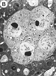
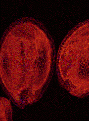
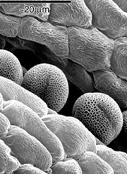
The Department has a modern electron microscope facility that is used by students, faculty, and staff. It is equipped with a Hitachi H-7650 transmission electron microscope, and a Hitachi S-4800 cold cathode field emission scanning electron microscope with a Bruker energy-dispersive spectrometry (EDS) system for elemental analysis. Ultramicrotomes, critical point dryers, vacuum evaporators, sputter coaters and fully equipped darkrooms are also available for use. Formal courses in scanning and transmission electron microscopy provide training to both graduate and undergraduate students.
Equipment
Electron Microscopes
Hitachi H-7650 Transmission Electron Microscope (TEM)
- accelerating voltage to 120 kV in 20 kV increments
- film and digital image acquisition
- specimen holders: standard, dual, rotational (goniometer), low background (beryllium) current metering, and cryotransfer (see below)
Hitachi S-4800 Ultra High-Resolution Cold Cathode Field Emission Scanning Electron Microscope (FE-SEM)
- operable in secondary electron (SE), backscattered electron (BE), and scanning transmission electron (STEM) modes
- capable of simultaneous brightfield and darkfield STEM imaging
- digital image acquisition
- exceeds manufacturer’s guaranteed resolutions of 1 nm at 15 kV, 2 nm at 1 kV, and 1.4 nm at 1 kV with beam deceleration on site (standard gold on carbon test specimen)
Energy-dispersive X-ray Spectrometry (EDS)
Hitachi S-4800 FE-SEM is equipped with a Bruker Quantax EDS system
- thin window silicon drift detector (SDD) allows the detection of elements Boron and higher
- point analysis, line scan analysis and digital dot mapping capabilities
TEM Specimen Preparation
- Two RMC MT 7000 Ultramicrotomes
- Reichert Ultracut E Ultramicrotome
- glass knife makers (RMC, LKB)
- LKB Multiplate
SEM Specimen Preparation
- Critical point dryers (Balzers, Tousimis)
- Sputter coaters (SPI, CS Mini Coater, EMITECH K575x with carbon coating attachment)
- Vacuum evaporator (Edwards 306A)
Cryo Specimen Preparation
- RMC Propane Jet Freezer
- Leica AFS Freeze Substitution Apparatus
- RMC CR-21 attachment for RMC MT 7000 ultramicrotomes
Light Microscopes
- Leica TCS SP2 Confocal System installed on a Leica upright compound microscope. Microscope additionally equipped for DIC, phase contrast and epifluorescence microscopy.
- 2 lasers provide 5 excitation lines for confocal imaging (458 nm, 476 nm, 488 nm, 514 nm, and 543 nm)
- 3 PMT channels for confocal acquisition, one transmitted light PMT
- Equipped with a Leica FLEXACAM C1 for digital image capture in other modes
- Olympus BH-2 upright compound microscope equipped for epifluorescence microscopy with a 100 watt mercury burner and 3 filter cubes (Olympus B L0910, B L0911 and B L012)
- Olympus PM 10AD photomicrographic system for acquisition of 35 mm photographs with Olympus BH-2 microscope
Image Analysis, Storage and Output
- Fully equipped B&W darkroom (including condenser enlarger, point-source enlarger)
- Computers and software for image analysis (Photoshop, Quartz PCI, Metamorph)
- Epson Perfection V750 Pro scanner
- B&W and color laser printers
- Epson inkjet photo printer
Miscellaneous
- Wet lab for TEM and SEM specimen preparation including incubators, ovens, balances, stereo and light microscopes, centrifuges, microcentrifuge, pH meter, hot plates, stir plates, fume hoods, etc.


