The biophysics and biomedical imaging group is comprised of six faculty members interested in a wide array of topics such as the structure and dynamics of biological macromolecules, collective behavior of macromolecular and cellular systems, algorithm development for direct image reconstruction of cancerous tissues. The faculty members are joined in their endeavors by several postdoctoral research associates, and graduate and undergraduate students. More details can be obtained from web pages of individual groups or faculty members and from other areas of the Physics Department website.
Faculty Research
 Carol Hirschmugl
Carol Hirschmugl is developing a rapid chemical imaging technique using infrared imaging microscope coupled to an synchrotron source, which will be used to examine real-time biochemical changes in vivo.
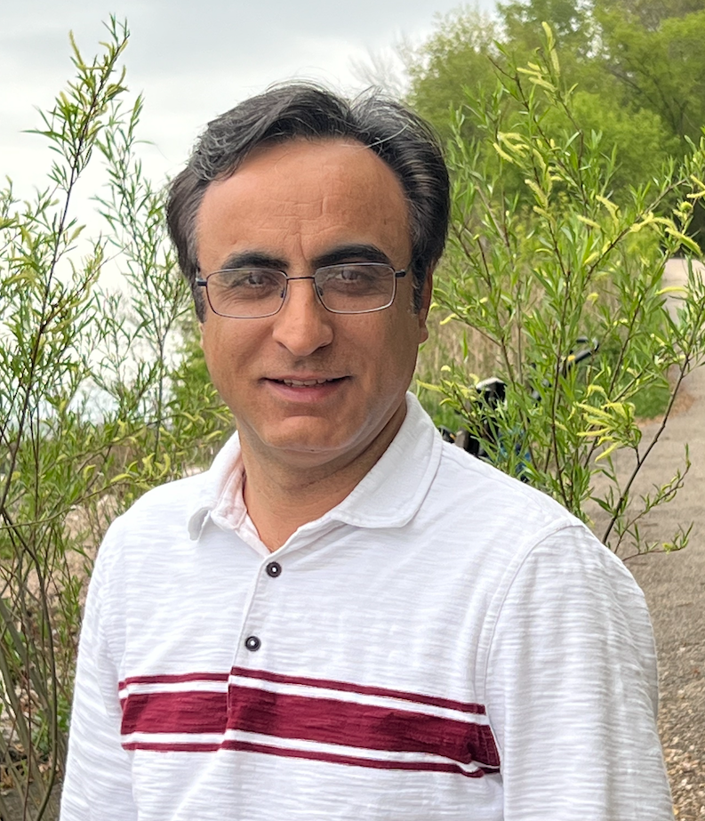 Ahmad Hosseinizadeh
Ahmad Hosseinizadeh’s research covers different topics in computational biophysics. His work centers around the development of novel methods for the analysis of large-scale data sets from biological molecules to understand the structure and function of these objects. A particular focus of his research is developing data-driven machine learning algorithms to extract the ultrafast structural dynamics of biomolecules at high spatial and temporal resolutions.
For more information: Hosseinizadeh’s Research Website
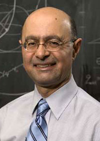 Abbas Ourmazd
Abbas Ourmazd’s research is aimed at developing innovative approaches for determining the structure and conformations of biological systems ranging from single molecules to whole cells at high resolution. The central hypothesis rests on new evidence that advanced algorithms stemming from Riemannian geometry, graph theory, and machine learning can be combined to forge a powerful new approach to biological structure determination in a way which circumvents the limits set by noise and radiation damage. These algorithms can be used with existing techniques, such as cryo-electron microscopy (cryo-EM), and emerging approaches exploiting the extreme brightness of X-ray Free Electron Lasers (XFELs). More generally, this work has important implications for tomography of faintly scattering, non-stationary objects at any length scale.
For more information: Ourmazd’s Research Website
 Ionel Popa
Ionel Popa’s research focuses on developing and implementing new techniques to study the mechano-chemistry of proteins. At the nanoscopic level force is a ubiquitous perturbation and many proteins have evolved to respond to mechanical stimuli. These proteins are generally segregated in multiple domains and play key roles in processes such as cellular mechano-transduction and tissue elasticity. The lab will use a new single molecule force spectroscopy technique based on magnetic tweezers to study the response of proteins to mechanical perturbations. This technique allows tethering of a single protein for more than a day and at forces similar to those encountered in vivo. Using this technique we aim to produce a detailed synthesis of the chemical and physical mechanisms that regulate proteins operating under mechanical force.
For more information: Popa’s Research Website
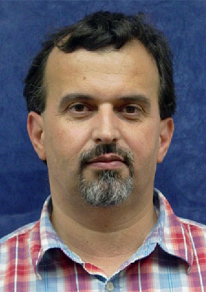 Valerică Raicu
Valerică Raicu is interested in experimental and theoretical aspects of the structure and dynamics of macromolecular and cellular systems in vivo. One of the main research themes in his lab is imaging of protein activity and interactions in living cells. This study involves novel approaches to laser-scanning fluorescence microscopy with spectral and temporal resolution, which is used in conjunction with a process of non-radiative energy transfer between fluorescent molecules, called Förster (or fluorescence) resonance energy transfer (FRET). The second major research theme in Prof. Raicu’s lab is the use of dielectric (or impedance) spectroscopy to probe the response to radiofrequency electrical fields of a material’s electrical polarization in order to determine its physical and structural characteristics at the microscopic level. This method is particularly suitable for characterization of biological cells individually dispersed in suspension or as part of supracellular associations, such as tissues and organs.
For more information: Raicu’s Research Website
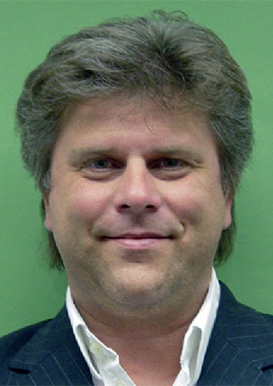 Marius Schmidt
Marius Schmidt’s research focuses on a unique method that unifies atomic structure with chemical kinetics: ultra-rapid, time-resolved X-ray structure analysis of proteins. This technique requires the brightest X-ray sources of the world, one of which is the close-by Advanced Photon Source at Argonne National Lab near Chicago. X-ray data collected there are four dimensional, with space and time as variables. With these data, chemical reactions can be observed directly in the protein crystals with a time-resolution of 100 pico-seconds and on the atomic length scale. The aim is to completely understand the catalyzed reaction including the atomic structures of the reaction intermediates and the corresponding kinetic mechanism. To analyze the large body of time-resolved X-ray data sophisticated and user-friendly software is required that is also developed by Professor Schmidt’s research group. In addition, proteins are produced, purified and crystallized for “de-novo” X-ray structure analyses.
For more information: Schmidt’s Research Website
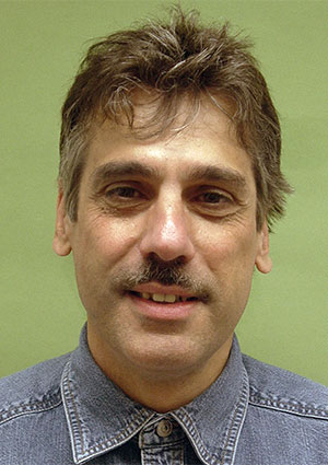 Peter Schwander
Peter Schwander’s research focuses on the structure and function of biological macromolecules by analyzing snapshots obtained with x-rays and electron microscopy. He is also a participant in BioXFEL, a Science and Technology Center (STC) established by the National Science Foundation (NSF) in 2013 consisting of eight U.S. research universities whose focus is addressing the fundamental questions in biology at the molecular level.