To study the swiftness of biology – the protein chemistry behind every life function – scientists need to see molecules changing and interacting in unimaginably rapid time increments – trillionths of a second or shorter.
Imaging equipment with that kind of speed was finally tested last year at the European X-ray Free-Electron Laser, or EuXFEL. Now, a team of physicists from the University of Wisconsin-Milwaukee has completed the facility’s first molecular movie, or “mapping,” of the ultrafast movement of proteins.
With this capability, scientists can watch how proteins do their jobs properly – or how their shape-changing goes awry, causing disease.
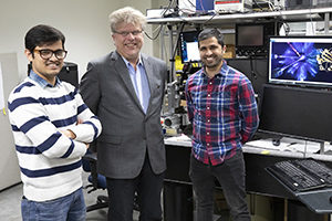
“Creating maps of a protein’s physical functioning opens the door to answering much bigger biological questions,” said Marius Schmidt, a UWM professor of physics who designed the experiment. “You could say that the EuXFEL can now be looked on as a tool that helps to save lives.”
Their findings marked a new age of protein research that enables enzymes involved in disease to be observed in real time for meaningful durations in unprecedented clarity. The paper is published online today in the journal Nature Methods.
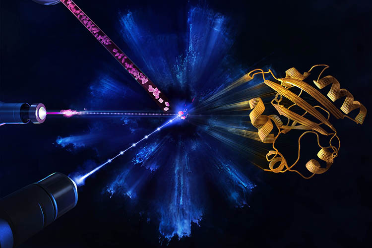
The EuXFEL produces intense X-rays in extremely short pulses at a megahertz rate – a million pulses a second. The rays are aimed at crystals containing proteins, in a method called X-ray crystallography. When a crystal is hit by the X-ray pulse, it diffracts the beam, scattering in a certain pattern that reveals where the atoms are and producing a “snapshot.”
The rapid-fire X-ray pulses produce 2D snapshots of each pattern from hundreds of thousands of angles where the beam lands on the crystal. Those are mathematically reconstructed into moving 3D images that show changes in the arrangement of atoms over time.
A new level
The European XFEL, which opened last year, has taken this atom-mapping to a new level. Extremely powerful bursts contain X-ray pulses at a quadrillionth of a second, in “bursts” that occur at 100 millisecond intervals.
Schmidt’s experiment began with a flash of blue, visible light that induced a chemical reaction inside the protein crystal, followed immediately by a burst of intense X-rays in megahertz pulses that produce the “snapshots.”
It’s an experiment he first staged in 2014 at the U.S. Department of Energy’s SLAC National Accelerator Laboratory in California. There, he and his students were able to document atomic changes in their protein samples for the first time at an XFEL.
Subsequently, in 2016, they were able to map the rearrangement of atoms in the range of time proteins take to change their shapes – quadrillionths of a second (femtoseconds) up to 3 trillionths of a second (picoseconds). In a picosecond, which is a trillionth of a second, light travels the length of the period at the end of this sentence.
Previous time-resolved crystallography on their photoreactive protein had already been completedusing other X-ray sources capable of imaging time scales larger than 100 picoseconds, leaving a gap of uncharted time between 3 and 100 picoseconds that the scientists were able to fill using the EuXFEL.
Much quicker data gathering
The exceptional brightness of the laser and the megahertz X-ray pulse rate allowed them to gather data much quicker and with greater resolution and over longer time frames.
Schmidt describes EuXFEL as “a machine of superlatives.” The largest XFEL in the world, it is 3 kilometers long, spanning the distance between the German federal states of Hamburg and Schleswig-Holstein. Superconductive technology is used to accelerate high-energy electrons, which generates the X-rays.
Schmidt, a biophysicist who has participated in more than 30 XFEL imaging projects to date, offered a taste of the medical potential of enhanced crystallography with the XFEL: Using this method, he has witnessed how multiple proteins work together, how enzymes responsible for antibiotic resistance disable a drug and how proteins change their shape in order to absorb light and enable sight.
Doctoral student Suraj Pandey, who came to UWM from his native Nepal, is first author on the paper. He now has experience with technology that few people in the world can claim, at least for now. He said he was not sure what to expect going into the experiment.
Pandey’s role was to analyze the data and calculate the maps of structural change. Of the millions of X-ray pulses that XFELs deliver, the majority don’t hit a target at all. In fact, only 1% to 2% diffract off a protein crystal, while the remaining pulses produce “noise” that must be removed from the data.
The team had other worries too, he said. It took months for Pandey to grow the protein required to produce the experiment’s crystals, but during their transport to Germany, the 5 grams of frozen protein was detained in customs for several days, during which some of it melted.