Development stage

  1. Lead Identificaiton
  2. In Vitro Efficacy

Health Condition & Disease

In the past, antiviral drug discovery focused almost exclusively on viruses that cause chronic, life-long, diseases, like AIDS, herpes and hepatitis.  The COVID-19 pandemic has revealed that this was a tragic mistake. Potent antivirals, like those discovered many years ago for the related SARS and MERS coronaviruses, were never developed for clinical use.  Millions of lives could have been saved if these compounds had been developed into drugs.

Direct-acting antivirals (DAAs) are defined as those that specifically inhibit viral enzymes that are needed for virus replication. Examples include the herpes drug acyclovir, AZT, the first drug approved to treat AIDS, and the hepatitis C drug sofosbuvir.  The term was coined to differentiate DAAs from other antiviral drugs, like interferon and ribavirin, which function mainly by modulating the host immune system.

For over 20 years, the Frick lab worked with numerous other labs and companies to develop DAAs to treat hepatitis C.  Many of these drugs are now used routinely in the clinic to cure chronic hepatitis C, which used to kill tens of thousands of Americans each year. With DAAs hepatitis C can now be cured with a single daily pill taken for eight weeks, with few if any side effects.  The goal now is to adapt these DAAs for use with other viruses, like SARS-CoV-2, the virus that causes COVID-19.

Drug Target Identification

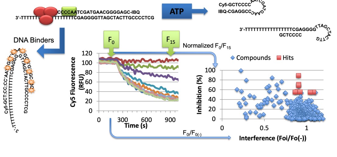
Most hepatitis C virus DAAs were discovered by biochemists using recombinant purified proteins because, unlike other viruses, HCV cannot be easily grown in the laboratory.  To facilitate these efforts, scientists in the Frick lab, isolated viral proteins and designed assays suitable for high-throughput screening. These enzymes include viral polymerases, proteases, primases, and helicases.  One example is an assay that can be used to discover viral helicase inhibitors and simultaneously differentiate them from toxic compounds that simply block viruses by binding nucleic acids (Fig. 1).

Scientific schematic of a DNA-based fluorescence assay workflow. DNA strands with labeled binders and ATP are illustrated at the top and left. A central line graph shows Cy5 fluorescence (RFU) decreasing over time with multiple colored traces labeled F₀ and F₁₅. On the right, a scatter plot displays inhibition (%) versus interference (F₀i/F₀(−)), with blue diamonds representing compounds and red squares representing hits. The figure summarizes screening of compounds for DNA-binding activity using fluorescence measurements.
Figure 1.  The Molecular Beacon-based Helicase Assay (MBHA).  Helicases are molecular motors that re-arrange nucleic acids in reactions fueled by ATP.  By using DNA that forms hairpins, an MBHA can be used to simultaneously differentiate compounds that inhibit helicases from compounds that might block helicases simply by binding the double helix. DNA binding agents are typically toxic and less promising drug candidates.  For details see Frick Lab papers in Biotechniques and Methods in Enzymology.

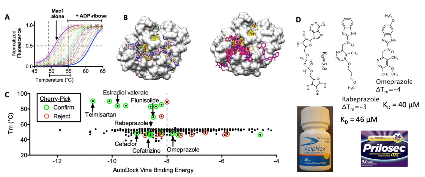
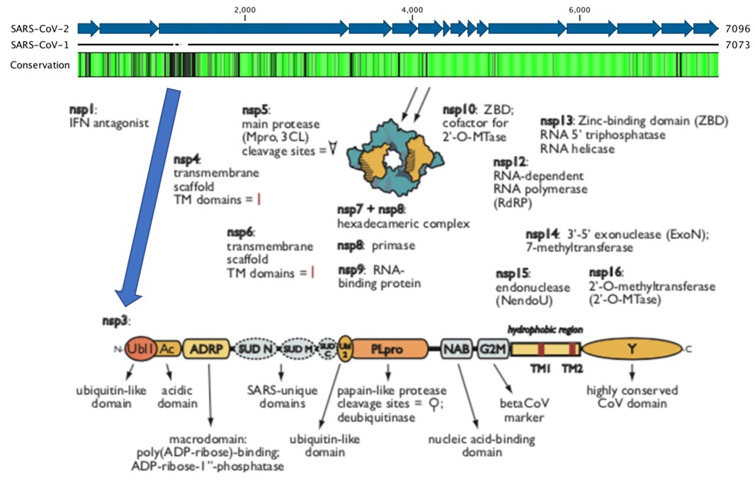
Because we desperately need DAAs to treat COVID-19, beginning in January 2020, the Frick Lab began isolating proteins from SARS-CoV-2.  The 29,900 nucleotide SARS-CoV-2 RNA genome encodes many potential DAA targets, most of which are encoded by the rep1ab open-reading frame.  The Frick lab has had most success with the multifunctional 945 amino-acid-long nsp3, which is tethered to the ER with two ubiquitin-like domains, two papain-like protease domains, three macrodomains (Mac1, Mac2, and Mac3), a nucleic-acid-binding domain, and a hypervariable region (Fig. 2).
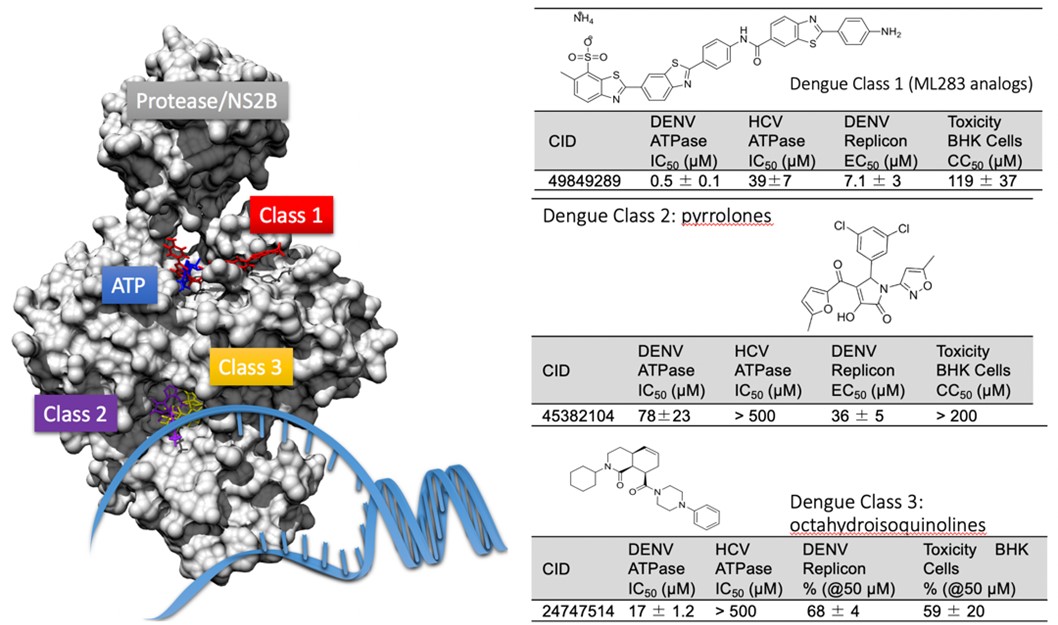
Figure showing a comparative genomic and functional map of SARS-CoV-2 and SARS-CoV-1 non-structural proteins (nsps). The top panel displays genome conservation between SARS-CoV-2 (7,096 nucleotides) and SARS-CoV-1 (7,073 nucleotides), with a color-coded conservation bar showing high similarity (green) and divergent regions (black). The middle panel annotates nsps 1 and 4–16 with their functions: nsp1 (IFN antagonist), nsp4 and nsp6 (transmembrane scaffolds), nsp5 (main protease/Mpro/3CL), nsp7+nsp8 (hexadecameric complex, illustrated as a teal and gold molecular structure), nsp8 (primase), nsp9 (RNA-binding protein), nsp10 (zinc-binding domain, cofactor for 2'-O-MTase), nsp12 (RNA-dependent RNA polymerase/RdRP), nsp13 (zinc-binding domain, RNA 5' triphosphatase, RNA helicase), nsp14 (3'-5' exonuclease/ExoN; 7-methyltransferase), nsp15 (endonuclease/NendoU), and nsp16 (2'-O-methyltransferase). The bottom panel provides a detailed domain map of nsp3, showing domains from N- to C-terminus: Ubl1 (ubiquitin-like), Ac (acidic), ADRP (macrodomain: poly-ADP-ribose-binding, ADP-ribose-1''-phosphatase), SUD-N, SUD-M, SUD-C/Ubl2 (SARS-unique and ubiquitin-like domains), PLpro (papain-like protease, deubiquitinase), NAB (nucleic acid-binding), G2M (betaCoV marker), a hydrophobic region with TM1 and TM2 transmembrane domains, and Y (highly conserved CoV domain).
Figure 2. Attractive SARS-CoV-2 DAA targets.  The above plot shows amino acids conserved (green) between SARS-CoV-2 and related viruses in the 1ab reading frame.  The 16 mature nonstructural proteins (nsp) are shown with arrows with functions noted below. Nsp3 is highlighted. The figure is adapted from Fields Virology (6th edition).

In Vitro Efficacy

After DAAs were approved and became the standard of care to treat hepatitis C, the Frick Lab began testing DAAs against similar viruses with RNA genomes like Dengue virus, West Nile virus, Zika virus and most recently SARS-CoV-2.  Many were potent enzyme inhibitors and blocked virus replication (Figs. 3, 4).  We now seek to develop this leads into antiviral drugs.

Figure illustrating three classes of Dengue virus inhibitors and their binding sites on the DENV NS3 helicase/NS2B protease complex. The left panel shows a molecular surface rendering of the protein (gray) with the Protease/NS2B region labeled in white at the top, and three distinct inhibitor binding sites highlighted: Class 1 (red label, near the ATP-binding site), Class 2 (purple label, lower left), and Class 3 (gold label, center). The ATP binding site is labeled in blue. A double-stranded RNA helix (blue) is shown threading through the base of the protein.
The right panel presents data for each inhibitor class:
Dengue Class 1 (ML283 analogs): Chemical structure of a bis-benzimidazole sulfonamide compound (CID 49849289). Data table shows DENV ATPase IC₅₀ = 0.5 ± 0.1 µM, HCV ATPase IC₅₀ = 39 ± 7 µM, DENV Replicon EC₅₀ = 7.1 ± 3 µM, Toxicity BHK Cells CC₅₀ = 119 ± 37 µM.
Dengue Class 2 (pyrrolones): Chemical structure of a dichlorophenyl pyrrolone compound (CID 45382104). Data table shows DENV ATPase IC₅₀ = 78 ± 23 µM, HCV ATPase IC₅₀ > 500 µM, DENV Replicon EC₅₀ = 36 ± 5 µM, Toxicity BHK Cells CC₅₀ > 200 µM.
Dengue Class 3 (octahydroisoquinolines): Chemical structure of a piperazine-containing octahydroisoquinoline compound (CID 24747514). Data table shows DENV ATPase IC₅₀ = 17 ± 1.2 µM, HCV ATPase IC₅₀ > 500 µM, DENV Replicon inhibition = 68 ± 4% at 50 µM, Toxicity BHK Cells = 59 ± 20% at 50 µM.
Figure 3. DAAs targeting the helicase encoded by Dengue virus.  The structure on the left is the Dengue virus NS2B/NS3 complex showing the binding sites for RNA, ATP and the various classes of DAAs discovered in screening campaigns.  Tables on the right show chemical structures and potencies in vitro and in cell based (replicon) assays and the PubChem compound identification (CID) number.  For more information see our paper in ACS Infectious Diseases.
Four-panel figure showing the identification of Mac1 macrodomain inhibitor leads using thermal shift assay, molecular docking, and binding validation. Panel A: Line graph showing normalized fluorescence (y-axis, 0 to 1.0) versus temperature (x-axis, 45–65°C) from a thermal shift assay. Two reference curves are shown: 'Mac1 alone' (blue, leftmost) and '+ ADP-ribose' (purple, rightmost), with multiple colored curves from compound screening plotted in between. A horizontal dashed line at 0.5 fluorescence marks the melting temperature (Tm). Panel B: Two molecular surface renderings (gray) of the Mac1 protein showing docked compound poses. The left image displays a diverse set of multicolored docked ligands clustered at the binding site (yellow region). The right image highlights a subset of top-scoring compounds shown in magenta/pink poses at the same binding site. Panel C: Scatter plot of Tm (°C, y-axis, 20–100°C) versus AutoDock Vina Binding Energy (x-axis, −12 to −4) for screened compounds. Green circled dots indicate confirmed hits; red circled dots indicate rejected compounds. Several compounds are labeled with arrows: Estradiol valerate, Flunisolide, Telmisartan, Rabeprazole, Cefaclor, Cefatrizine, and Omeprazole. A vertical dashed line is drawn near −8 kcal/mol. Panel D: Chemical structures and binding data for two confirmed lead compounds. Rabeprazole (proton pump inhibitor; ΔTm = −3, KD = 46 µM) and Omeprazole (proton pump inhibitor; ΔTm = −4, KD = 40 µM) are shown with their chemical structures alongside photographs of their commercial drug products: AcipHex (rabeprazole sodium, 20 mg delayed-release tablets) and Prilosec OTC (omeprazole, 42 tablets).
Figure 4. Identification of FDA approved Drugs that might be templates to design DAAs for SARS-CoV-2.  (A) Design of an HTS-compatible ADP-ribose binding assays to detect inhibitors of the Mac1 domain of SARS-CoV-2 nsp3. (B) Virtual screens using AutoDock Vina to predict where compounds might bind Mac1.  (C) When libraries are screened with these assays desirable drug candidates should bind with lowest free energy and highest Tm.  (D) Two similar FDA-approved proton pump inhibitors that bind Mac1.  Note that others have shown that omeprazole is a modest antiviral in cell culture.  Clinical trials with these drugs have been initiated.  For more information see our paper in SLAS Discovery.