The bacteria that cause tuberculosis are particularly difficult to treat because they are often antibiotic resistant. In fact, the lung disease, which spreads through coughs or sneezes, now kills more people worldwide than any other infectious agent, according to the World Health Organization.
But in order to design drugs that foil resistance, scientists first have to know the how the bacterium disables the antibiotic at the atomic level.
In experiments led by UWM Physics Professor Marius Schmidt, a team of researchers has watched the process in action for the first time using an X-ray free-electron laser, or XFEL, at the Department of Energy’s SLAC National Accelerator Laboratory.
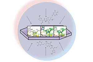
In the experiment, Schmidt and his colleagues mixed an antibiotic with an enzyme, a class of proteins, that TB bacteria produce to protect themselves. Then scientists watched in real time as the enzyme attacked the antibiotic molecule and burst one of its chemical bonds.
“This proof-of-concept study shows that we’re able to see the shape and intermediate stages of the molecules during the process,” Schmidt said. “After decades of trying other techniques in the field of crystallography, the technology is (now) here.”
The results of the experiment were published May 31 in BMC Biology.
In the past, the only way to image the atomic structure of molecules and proteins in three dimensions was with X-ray crystallography: X-rays are shot at crystalized proteins, which diffract the light and create patterns, or “snapshots,” of dots the way shaking a paintbrush sprays drops on a wall. Each of these snapshots reveals the protein’s atomic structure at a single point in time.
Using SLAC’s light source, XFEL imaging turbocharges that method, allowing images to be taken in extremely short time increments.
For this experiment, researchers started the reaction by mixing the antibiotic and bacteria just fractions of a second before the sample was hit by the XFEL’s X-ray ultra-bright pulses. Snapshots were taken 30 milliseconds to two seconds after the reaction began.
Then scientists stitched together the millions of snapshots during the reaction, creating a map that shows how the atoms of the enzyme and antibiotic molecule are rearranged as they interact at room temperature.
XFELs have such intense beams that they can capture diffraction patterns from much tinier crystals, a millionth of a meter across or less, Schmidt said, so the antibiotic could get to the enzyme before bombardment with X-rays.
“For structural biologists, this is how we learn exactly how biology functions,” says Mark Hunter, staff scientist at SLAC and co-author on the study. “We decipher a molecule’s structure at a certain point in time, and it gives us a better idea of how the molecule works.”
Schmidt previously has used XFEL imaging to investigate how a photo-reactive protein involved in light perceptionchanges its shapeas it interacts with a photon, a particle of light.
Next the researchers intend to apply this method to additional pharmacologically important enzymes, with an eye on providing new targets for drug development.
Schmidt collaborated with two other UWM physicists, Abbas Ourmazd and Peter Schwander. UWM graduate students Suraj Pandey and Ishwor Poudyal, research associate Christopher Kupitz and Jacob Verburgt, an undergraduate intern from the Milwaukee School of Engineering, also were involved in the work.
Schmidt and the UWM team are members of a BioXFEL Science and Technology Center backed by the National Science Foundation, which formed the collaborative framework for this work. In addition to UWM and SLAC, the project included scientists from six other universities, and Deutsches Elektronen-Synchrotron, the Hamburg Center for Ultrafast Imaging, the Lawrence Livermore National Laboratory, Max Planck Institute for Biochemistry, GlaxoSmithKline and 4Marbles Inc.