Researchers in UWM’s Department of Physics have recently been awarded special funding that will allow them to probe different aspects of SARS-coV-2, the virus that causes COVID-19, using molecular imaging techniques. Their work, chosen on merit, is funded by two grants from the National Science Foundation that support exploratory research designated for an urgent need.
What affects the coronavirus’s severity?
The proteins in nearly all biological forms, including the current coronavirus, undergo “conformational” changes – a morphing of their atomic shape as they respond to their environment. Scientists think seeing these changes in action will help clarify what happens when SARS-coV-2 and its later mutations turn virulent.
Obtaining molecular pictures over split-second intervals requires advanced imaging techniques – methods that, in turn, rely on creative data science.
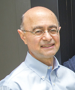
In this project, machine-learning algorithms developed by a group led by Distinguished Professor Abbas Ourmazd will allow scientists to see relevant proteins in the virus change both by themselves and also while interacting with antibodies produced by the body’s immune system.
The goal is to reveal insight into the way the virus functions and, ultimately, provide a foundation for the development of therapeutic strategies against coronaviruses.
How the virus becomes infectious
SARS-coV-2 can’t reproduce on its own. It has to first infect a cell and then use the cell’s machinery to replicate.
The 3CLpro protein of the virus acts as “molecular scissors,” custom-cutting virus proteins, so that they can form new virus copies. Like real scissors, 3CLpro accomplishes that by rearranging its atomic structure. Scientists want to use advanced molecular imaging equipment, called X-ray Free Electron Lasers, or XFELs, to film each atomic component as the protein does its job.
Results will aid the design and discovery of new drugs that would block the 3CLpro protein so that new virus particles cannot assemble and become infectious.
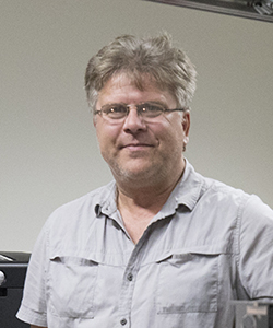
Professor Marius Schmidt and his students will introduce small compounds that interrupt the protein’s work at the XFEL facilities at Stanford University and in Hamburg, Germany. The XFEL can capture a molecular picture every 100 femtoseconds (a quadrillionth of a second), which Schmidt and the other researchers will piece together to form a slow-motion movie of the protein changing in real time.
Associate Professor Ionel Popa will help in the production and crystallization of the 3CLpro.