A pharmaceutical tablet may look simple from the outside. But what if that tablet contains several medications, arranged in different regions and designed to release at different rates?
As the manufacturing of pharmaceuticals becomes more complex, knowing what is happening inside the tablet is critical. Yet current quality-control imaging techniques are inadequate because the ingredients absorb and scatter light so strongly.
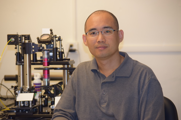
Yongjin Sung, an associate professor of mechanical and biomedical engineering, is developing a new tool that could allow researchers to look inside these tablets without cutting them open or destroying them.
In his $518,000 project, funded by the National Science Foundation, he and collaborators at UC-San Francisco (UCSF) will develop imaging that can determine what chemicals are present inside a specimen, where they are located, and how deeply they extend.
Benefits emerging 3D-printed pharmaceuticals
The work is particularly relevant to 3D-printed pharmaceuticals, an emerging method of forming tablets that contain several medications or deliver them at different rates. Unlike conventional pills, 3D-printed tablets can contain multiple active ingredients arranged in carefully controlled patterns that minimize drug interactions and side effects.
“The ingredients are formulated so that the various active components, which act in different ways, remain separate from one another,” Sung said.
The goal is to create an instrument that can confirm whether those ingredients actually ended up where they are supposed to be.
Combining techniques
Sung’s approach blends optical imaging with computational methods to overcome the challenges.
The project builds on diffuse optical tomography, an imaging technique that has been widely investigated for medical imaging of the human body. Because biological tissue strongly scatters light, diffuse optical tomography uses measurements of light that has traveled through tissue to reconstruct information about structures inside the body.
Pharmaceutical tablets present a different and more difficult challenge because they can be both strongly scattering and strongly absorbing. It is not yet known how well diffuse optical tomography can be applied to such materials, Sung said.
The team will build a new imaging system operating in the extended short-wave infrared range, a region of light invisible to the human eye that contains rich information about the chemical composition of materials.
Sung will work with Professors Youngho Seo and Grant Gullberg, medical imaging experts at UCSF, to develop computational methods for recovering chemical information from deep within these specimens.
Instead of looking like a photograph, the result would be more like a 3D chemical map.
The work builds on Sung’s broader research in advanced imaging and measurement techniques for biomedical, semiconductor and pharmaceutical applications.
His previous work includes snapshot optical tomography – a method that captures a 3D image of a specimen in a single shot – and advanced X-ray and PET imaging technologies.