What happens to the human retina at the cellular level when it’s exposed to the hyperglycemic conditions of diabetes? No known imaging technique has been able to show such biochemical changes – until now.
Located in the back of the eye, the retina’s layers of cells convert incoming light into a signal that the brain processes. Damage to the retina from diabetes leads to a condition called diabetic retinopathy, a leading cause of blindness.
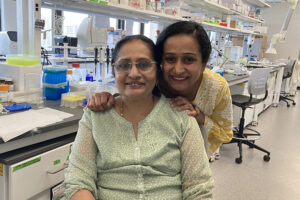
A research team led by the University of Wisconsin-Milwaukee has revealed changes in the retinal cell layers that drive the progression of this disease.
Their unique imaging technique identified biomarkers of cellular damage occurring over a 10-month period in mice. Diabetic retinopathy affects multiple layers of the retina simultaneously.
“We know the basic progression of the disease,” said Carol Hirschmugl, UWM professor of physics. “But if we knew what was happening at the cellular level, it could lead to a more effective treatment or earlier diagnosis.”
Results showed that the retina’s photoreceptors – cells in the eye that react to photons and then signal the brain – are the most susceptible cells to the damage at the onset of the disease. At six weeks into the disease however, the chemistry of only one retinal layer showed changes, while the others appeared healthy.
The study, conducted with researchers at the McPherson Eye Research Institute at UW-Madison, confirms earlier research suggesting that retinal deterioration occurs at irregular intervals rather than steadily declining – and it uncovered that significant cell damage occurs within the first three months.
Imaging involved a tool Hirschmugl developed that concentrates the intense light of a synchrotron in the infrared wavelengths. Combined with a technique called Fourier transform infrared (FT-IR) imaging, the researchers recorded a molecular fingerprint for functional groups such as proteins, carbohydrates and lipids that can signal the development of retinal diseases.
“We were studying the combined chemical signature of everything in the samples,” Hirschmugl said. “Since we use all the colors at once, we can tell you about all things in one picture. We could also detect what was missing.”
In order to complete the imaging without the use of a synchrotron facility, the scientists used the previous synchrotron data to create computer algorithms that compensate for the reduced quality of the typical FT-IR measurements.
Now they have a platform for applying the imaging technique to study other diseases of the retina. And the platform has the potential to find damaged cells in other disease models.
From longitudinal studies, researchers found that the rate of cellular damage increased from six to 12 weeks of diabetes, and then decreased between three and six months, followed by a gradual increase by the 10th month of the disease. In fact, the greatest number of proteins, which are responsible for all cellular processes, were significantly damaged six months into the disease.
Why does the rate of deterioration plateau at three months?
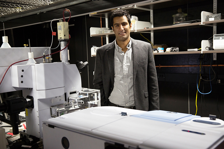
“We believe that these temporal variations in biomarkers correlate with the cells’ mechanisms that repair the damaged or degraded macro-molecules,” said Ebrahim Aboualizadeh, a postdoctoral research associate at the University of Rochester who was first author of the recent papers published in the journals Scientific Reports and Trends in Biochemical Sciences.
Ultimately, however, the rate of cell injury outpaces the body’s ability to repair the damage.
Hirschmugl has used the equipment she developed for the synchrotron in other applications, such as determining the presence of malaria parasites in blood samples as small as a single cell.