A better look at diabetic brains
Consistently high blood sugar levels caused by Type 2 diabetes can damage organs and blood vessels, and if the damage occurs in the brain, result in stroke and dementia.
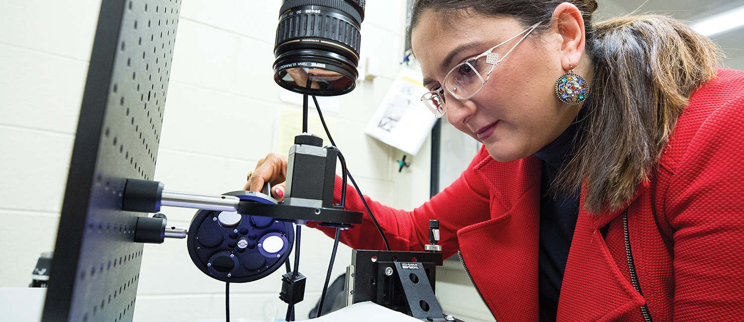
Medications help about 28 million Americans limit these risks. But UWM researchers believe more can be done. What if they could recognize subtle changes in a diabetic’s brain before there was irreversible harm? That’s the long-term goal of Mahsa Ranji and Ramin Pashaie, associate professors of electrical engineering in the College of Engineering & Applied Science.
The researchers are developing innovative imaging technology that detects tiny alterations in the brain’s blood flow and metabolism, which could eventually damage vessel or nerve cells. Their approach offers both visual and quantitative measurements of these changes, and Ranji says a detailed quantitative analysis provides much more objective results than those that come from qualitative image comparisons made with the human eye.
“Our hypothesis is that vascular and metabolic changes in the diabetic brain are linked,” Ranji says. “Imaging both processes over the same period of time in living tissue has never been done before.”
The project is a unique combination of its leaders’ expertise.
Pashaie has developed portable technology for tracking blood flow that can be customized for the brain and many other tissues. Ranji is an expert in fluorescence imaging and biomedical optics. It’s a type of imaging that makes use of fluorescence to image cells and living tissue at greater depth than other methods.
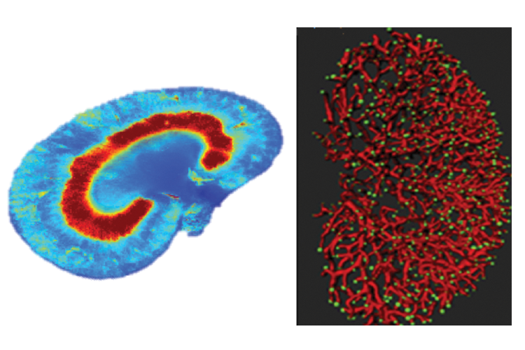
Scans focus on two proteins that help convert the chemical energy contained in blood sugar to energy that brain cells can use. Both proteins naturally emit absorbed light, which makes them ideal for fluorescent imaging.
Ranji measures the ratio of the two proteins as a marker of the tissue’s metabolic health. Abnormal values suggest that diabetes may disrupt the brain’s energy conversion.
Ranji notes that the work is in its early stages.
“If our approach detects a direct correlation between vascular and metabolic brain changes in mice, that would be a major finding,” Ranji says.
“Years down the road,” she adds, “it may help design new interventions at early stages of human diabetes to reduce the risk of complications, such as stroke or dementia.”