Nuclear imaging, such as positron emission tomography (PET), provides superior sensitivity and specificity. That is why PET is widely used in hospitals to detect and stage different types of cancers in the body.
However, the resolution of PET images is limited.
To overcome this, a group at Stanford University has developed a novel technique called radioluminescence microscopy (RLM) that also uses radioisotopes as tracers. But RLM can provide much higher resolution by directly detecting positrons, particles emitted by the radioisotopes, instead of annihilation gamma rays produced by the positrons.
RLM is the only technique that can measure the distribution of radioisotope uptake among single living cells, and it has led to many important discoveries.
The resolution of RLM, however, quickly drops as the sample thickness increases, which is common to microscopic imaging techniques detecting positrons. “That leaves scientists with no microscopic nuclear imaging technique with depth-resolving capability,” said Yongjin Sung, associate professor, mechanical and biomedical engineering.
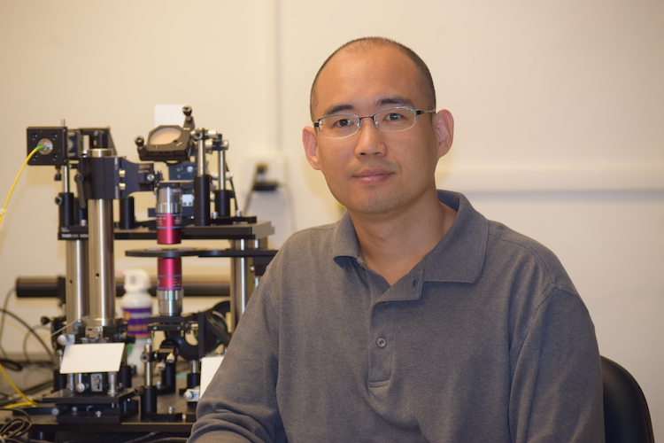
Sung and his colleagues have been awarded a $547,700 grant from the National Institutes of Health to add the depth dimension to RLM.
Ultra-quick and ultra-tiny
In this grant, Sung is collaborating with Guillem Pratx, a professor at Stanford University who has pioneered advances in RLM, and Youngho Seo and Grant Gullberg, professors at the University of California-San Francisco who are PET imaging experts.
The idea enabling the innovation is to capture the 3D shapes of positron trails and track the positrons back to the radiotracers.
“The task is similar to taking a 3D picture of lightning at a microscopic scale,” Sung said.
“The positron trails disappear within tens of microseconds. However, using a method called snapshot optical tomography, we can capture their 3D shapes with a single snapshot.”
Living cells from a specific patient
RLM imaging takes place outside the human body and can be applied to the cells taken from a specific patient.
“With RLM, we can test all different types of radiopharmaceuticals and find the best therapy for that individual patient,” Sung said.
With the improved resolution and depth-sectioning capability, he said, the 3D RLM technique will open new doors to microscopic nuclear imaging and allow for testing new radiotracers using living, multi-cellular organisms before translating to small animals and human patients.