The University of Wisconsin-Milwaukee (UWM) and partners at seven leading U.S. research institutions have landed a highly competitive $25 million grant from the National Science Foundation (NSF) to conduct work that could transform the way scientists study diseases and find new treatments.
The funding establishes an NSF Science and Technology Center (STC) that will use powerful X-ray lasers to reveal the structure of proteins and viruses, and the way they work. STCs are considered one of the most prestigious NSF awards, and only three were awarded in this four-year cycle.
The University at Buffalo is the lead institution, with UWM and Arizona State University playing key roles. Other partner institutions are Cornell University; Rice University; Stanford University; the University of California, Davis; and the University of California, San Francisco.
Next-generation crystallography
Proteins are behind nearly everything that happens in our bodies, and malfunctioning proteins are often the cause of disease, making them a prime target for therapeutic drugs. But drug discovery is often a process of trial and error.
That’s because for many proteins, one important piece of information is missing: their structure – how their atoms are arranged. And structure reveals function.
The award will allow UWM and partners to build on their pioneering work in determining the structure and function of proteins not amenable to existing techniques.
More important than finding the structure of proteins individually, the team wants to determine how they operate within the organism.
The most widely used method of imaging biological molecules currently involves forming a pure crystal and bombarding it with X-rays. The pattern of rays as they diffract off the sample reveals a kind of fingerprint of the atoms in the molecule. A mathematical computation uses this to deduce the locations of the atoms.
Fewer than 20 percent of proteins currently form crystals large enough for this crystallography technique, including the majority of proteins in the outer wall of cells.
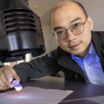
“Cell membrane proteins control the flow of information and material into and out of cells,” says Abbas Ourmazd, Distinguished Professor of Physics and Electrical Engineering, who leads the UWM team. “But they are notoriously difficult to crystallize – if it can be done at all, it takes a very long time.”
What small crystals can do

The first is an X-ray free-electron laser (XFEL), which produces X-ray light more than a billion times brighter than any made by existing equipment. Of the three that exist, the closest is at the Stanford Linear Accelerator Center (SLAC) in California.In their quest to image single proteins and viruses, researchers in the STC will rely on two secret weapons – one of which was developed at UWM.
It means crystallography can now be done with protein crystals a thousand times smaller than before – even those at the nanoscale. Since smaller crystals are much easier to form, it opens the door for seeing a vast array of proteins not amenable to standard approaches.
Beyond crystals
Imaging with the XFEL can take advantage of the intensity and unimaginably short flashes of light to produce snapshots of even a single tumbling protein molecule.
The process is then repeated over and over with other single proteins of the same kind, producing terabytes of data related to the millions of X-ray diffraction snapshots, each from a different unknown angle.
The team’s second trump card is a tool to analyze the mountains of data. Developed by Ourmazd, Schwander, UWM Distinguished Professor of Physics Dilano Saldin, Senior Scientist Russell Fung and UWM engineers Roshan D’Souza and Ali Dashti, such algorithms mine huge amounts of data to piece together a 3D image of the molecule.
“Imaging with this new kind of X-ray scattering can speed the process of determining protein structures from years to only days,” says Ourmazd. “Part of the reason can be attributed to the mathematical procedure we have developed.”
Proteins in action
The brightness of an XFEL flash may also allow scientists to “see” protein molecules in action for the first time. The idea is to capture a series of images over time, displaying the structural rearrangement taking place as a protein carries out a task.
“We hope to see, and perhaps even make movies of, ribosomes tapping out proteins in a cell, and photosynthesis in a plant,” says Ourmazd. “More important than finding the structure of proteins individually, we want to determine how they operate within the organism.”
Performing a “pump-and-probe” experiment, Associate Professor Marius Schmidt, the UWM team’s only experimentalist, will use a synchronized laser that emits visible light to stimulate a photo-reactive protein, followed by the X-ray pulse of the XFEL, which probes the progress of the protein during the reaction.
“When we shoot larger crystals with the laser there are not enough photons to get the excitation [structural change] going,” says Schmidt. “That’s the nanocrystal advantage.”
Very small crystals contain fewer molecules, requiring fewer photons to prompt the changes.
“The next part of the story is even cooler,” he says. “It’s possible that with this improved imaging method, we won’t need crystals at all.”
Beyond pictures
The UWM team has developed algorithms that mine huge amounts of imaging data to piece together a 3D view of molecules.
While Ourmazd’s work deals primarily with single proteins, Saldin’s research focuses on determining the structure of proteins from multiple copies randomly oriented in solution, which is closer to their state in a living organism.
“The theoretical team is developing techniques that may ultimately allow us to break the shackles of crystallography, using the XFEL,” he says.
Not only that, but his work with Schmidt aims to follow fast structural changes of proteins in solution, such as in the ubiquitous phenomenon of photosynthesis.
For this work, the XFEL can do the job, he believes, because the flashes of light it produces are so short, they can capture the equally lightning-quick molecular changes happening in a group of proteins.
Creating molecular movies and imaging proteins without crystals is still a long way off, says Ourmazd. “But the STC award demonstrates the tremendous potential of the proposed approaches for solving key problems in the life and energy sciences.”