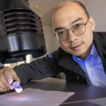
With a transformational method of imaging biomolecules that he helped to develop, Joachim Frank, who recently won the Nobel Prize in chemistry, found that molecules inside of human cells – the cellular machinery that powers our bodies – are in a constant state of motion.
He realized the potential of the imaging method, cryo-electron microscopy (Cryo-EM), after his lab created three-dimensional pictures of a biomolecule called a ribosome making proteins inside of a cell. The imaging has touched off a revolution that allows scientists to see, for the first time, “movies” of the basic processes of life that will benefit medicine.
“I knew then I could really contribute something to biology,” Frank said in a lecture he gave at UWM March 9. “I got resolutions that had never been achieved by anyone else.”
The Columbia University professor came at the invitation of Abbas Ourmazd, UWM distinguished professor of physics, who has published recent papers with Frank. This was the second Nobel laureate to visit UWM in less than six months. Bernard Feringa, who shared the 2016 Nobel Prize in chemistry, visited in September.
Frank shared the Nobel Prize in October 2017 with Richard Henderson of Cambridge University and Jacques Dubochet of University of Lausanne in Switzerland, for developing Cryo-EM. It allows scientists to view the movement of biological molecules using beams of electrons instead of light to take “snapshots” of samples after freezing them very quickly to preserve their natural shape.
Biomolecules change their atomic structure continuously as they accomplish cellular tasks like turning genes on and off, mediating chemical reactions, signaling other cells, and controlling cellular gateways. Before Cryo-EM, very few biomolecules could be imaged using other methods.
The imaging method produces multitudes of random, unsorted two-dimensional views that must be ordered and mathematically reconstructed. Frank created computational software that turned the blurry 2-D pictures taken with an electron microscope into detailed 3-D images.
In his lecture, which drew an audience of about 250, he traced the steps involved in perfecting Cryo-EM and also described recent collaborative work between his lab at Columbia University and the Ourmazd group.
Their work solved a specific problem: When the dataset is so huge, the shots all begin to look the same. Ourmazd’s group was able to create a new generation of powerful algorithms that bring out clarity at split-second intervals. Together, the scientists have made 3-D movies of some of the smallest units of life acting in ultra-short-lived states.
Frank said he credits interdisciplinary research and teamwork with the development of the technology. “I never thought, during all those years, that we would ever get this,” he said. “It was a dream come true.”