Proteins show their true colors
Proteins carry out every process necessary for life, and they take their instructions from the body’s genes. Learning more about how they work would help demystify gene mutations and provide new targets for pharmaceutical research.
The problem is that most proteins are notoriously difficult to image. Proteins often work in combinations called complexes, and current imaging methods yield only limited information about them. But new technology at UWM’s Small Business Collaboratory lets researchers observe proteins interacting within living cells, something they haven’t been able to do before.
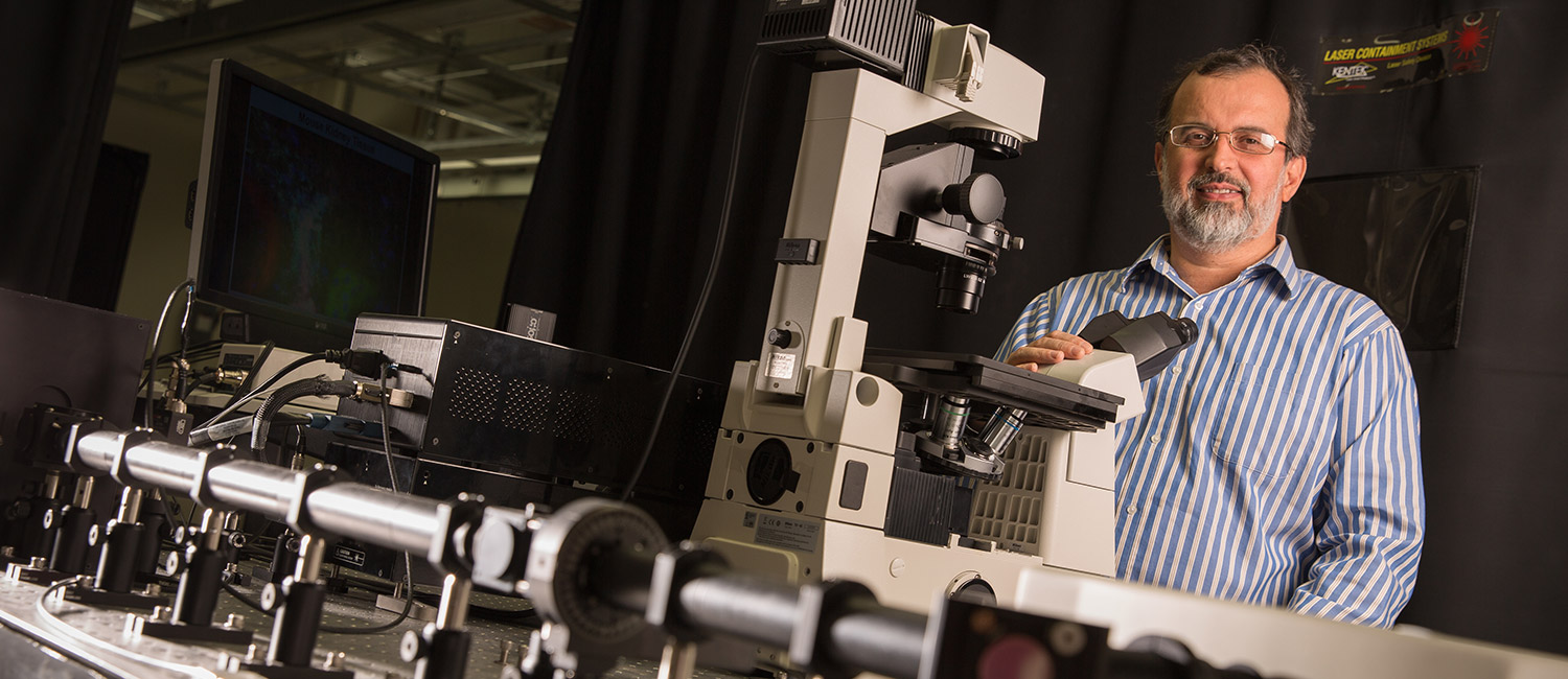
Constructed by physics professor Valerica Raicu, the equipment consists of a custom multiphoton microscope with laser attachments. It provides a color-coded view of how proteins and their complexes are distributed in a cell. “Think of the protein molecule as a car made of many parts,” Raicu says. “Rather than showing you just the parts, I can show you how they all are assembled to make the car.”
In two-dimensional images, each protein is tagged with a different fluorescent color. A laser “activates” the color of only one protein. But when the laser-excited protein interacts with another protein that’s unstimulated by a laser, it “gives up” its color. Then, only the second protein’s color is visible. The microscope can provide 3-D images by knitting together one thin slice of the image at a time. It also can show more than one color at a time, so researchers can see multiple interactions.
The new instrument is already rewriting some long-held beliefs about proteins that were based on how they interacted in lab conditions instead of in live tissue.
Paul Park, an assistant professor at Case Western Reserve University, has used it to watch live proteins in a human retina. A pigmented protein in the retina sparks biochemical reactions that trigger sight. Park wanted to see how that protein was arranged during the process. Ultimately, he wants to discover how certain gene mutations lead to impaired vision.
Raicu built the first iteration of the equipment with a Major Research Instrumentation grant from the National Science Foundation in 2011. Then, with more NSF support, he opened the collaboratory in 2014 so researchers from businesses, startups and other institutions could use the equipment.
With feedback from users and a new MRI grant, Raicu is now building an improved version of the microscope.
MRI grants are very competitive, and a single institution can only apply for three of them each year. In addition to Raicu, UWM physics researcher Patrick Brady and faculty in the Department of Chemistry and Biochemistry earned MRI grants. Winning three in one year is a rare feat for an institution, and it netted $1.7 million for equipment upgrades.